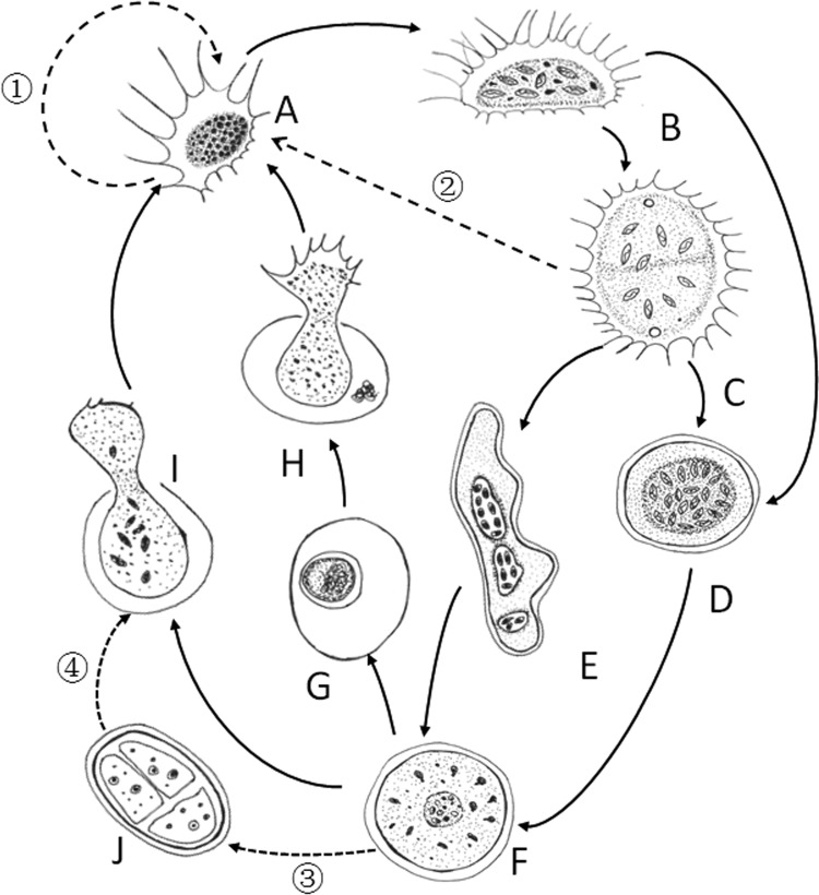
FIG 9.
Diagram showing transformations in gross morphology in the possible life cycle of V. algivore. (A) Trophozoite. (B) Trophozoite with ingested algal cells. (C) Plasmodia formed by fusion of two trophozoites. (D) Round digestive cyst in an early stage. (E) Irregularly shaped digestive cyst transformed from plasmodium. (F) Digestive cyst in a late stage. (G) Resting cyst. (H) Excystment of a young trophozoite from the resting cyst. (I) Excystment of young trophozoites from the digestive cyst. (J) Cell division inside the cyst. The solid arrows indicated that the transformations were observed in this study, while the dashed arrows indicate that the transformations were not observed in our study but probably existed. As is shown, cell division may take place in the trophozoite stage (A, 1), the plasmodium stage (C, 2), or the digestive cyst stage (J, 3 and 4) or during excystation (I). Due to the dense matrix of the central cytoplasm, it was not usually possible to observe the distinct organelles, such as nuclei and contractile vacuoles.

