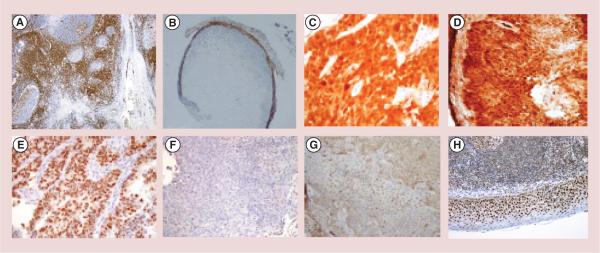
Figure 5. Immunohistochemical analyses of immune system interaction with the antibodies treated CasKi tumors.

(A) Positive control for CD3 positive cells (tonsil); (B) lymphocytic invasion with CD3 positive cells of the tumors treated with unlabeled TVG701Y monoclonal antibodies (mAb) to E7. CD3 positive cells stain brown; (C) positive control stained for p16 (metastatic squamous cell carcinoma); (D) tumor treated with unlabeled C1P5 mAb stained positive for p16; (E) positive control for p53 (endometrial intraepithelial serous carcinoma); (F) tumor treated with unlabeled C1P5 mAb stained negative for p53; (G) tumor treated with unlabeled TVG701Y mAb stained positive for retinoblastoma; (H) positive control for retinoblastoma (tonsil).
