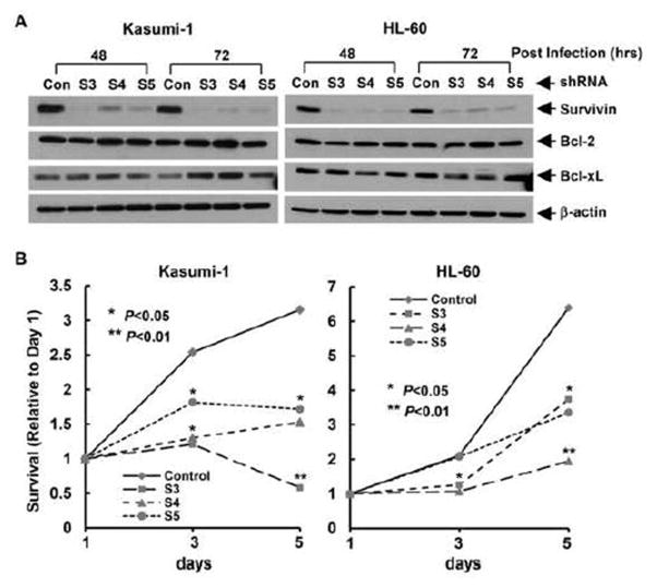
Figure 2. Specific knockdown of survivin expression inhibits cell proliferation.
Kasumi-1 and HL-60 cells infected with lentivirus containing either ConshRNA (Con) or SurshRNA (S3, S4, and S5) were subjected to the following experiments. A, After 48 hr or 72 hr lentiviral infection, cells were collected and subjected to Western blot analyses with specific antibody directed against survivin, Bcl-2, Bcl-xL, or β-actin. B, After 48 hr lentiviral infection, cells were plated onto 96-well plates at the density of 8000 cell/well, MTS assays were performed to examine cell growth. All wells were read at 490nM with a microplate reader on Day 1, 3, and 5. Reading of Day 1 was set as control. Curves show the relative proliferation rates of cells.

