Fig. 1.

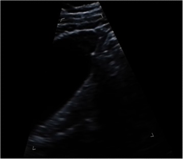
US image of fundal localized type GB adenomyomatousis. Localized thickness of GB wall in form of triangle at the fundus with small anechoic areas was detected

US image of fundal localized type GB adenomyomatousis. Localized thickness of GB wall in form of triangle at the fundus with small anechoic areas was detected