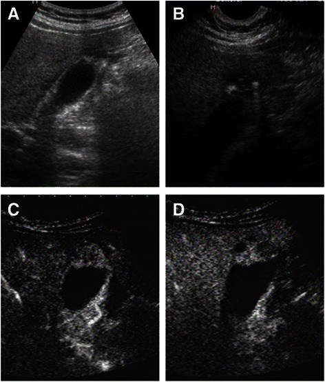
Fig. 3.

Image of US and CEUS. a The fundus of GB wall was thickened and the GB wall was obscure. b The intramural echogenic foci were detected by high frequency transducer. c CEUS—arterial phase (22 s) —heterogeneous hyper-enhancement and wall was intact. d CEUS—venous phase (34 s) the anechoic spaces were more clear
