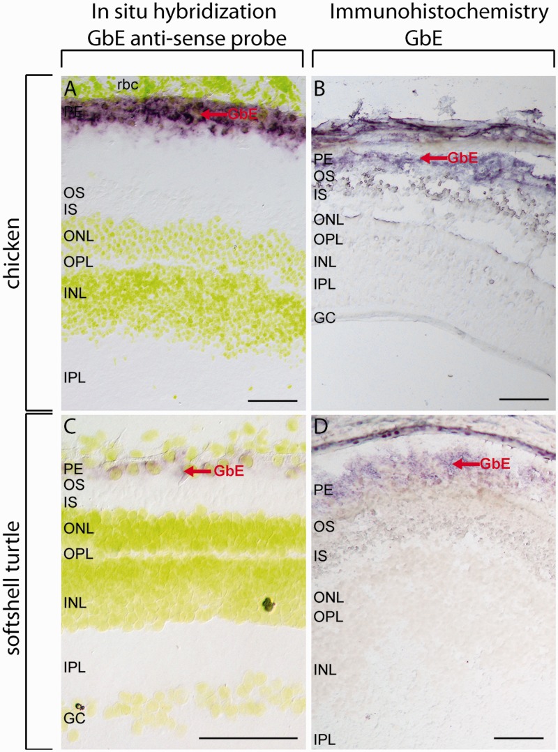
Fig. 5.—
Localization of GbE in the retina of softshell turtle and chicken. ISH was carried out with a species-specific antisense probe to detect the GbE mRNA (A, C); for IHC, a specific GbE-peptide antibody was used to detect the GbE protein (B, D). Both GbE mRNA and protein were detected in retinal PE, indicated by red arrows. In ISH, the nuclei were stained with Hoechst dye 33258, shown in yellow. Negative controls with sense probes (ISH) and omitted first antibody (IHC) are shown in supplementary fig. S4, Supplementary Material online. Scale bar = 100 µm. PE: pigment epithelium, OS: outer segments of the photoreceptor cells, IS: inner segments of the photoreceptor cells, ONL: outer nuclear layer, INL: inner nuclear layer, IPL: inner plexiform layer, GC: ganglion cells.

