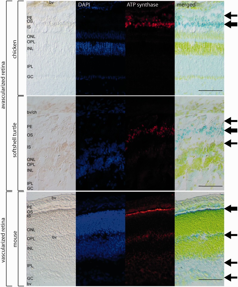
Fig. 7.—
Immunofluorescence of ATP synthase beta in avascular retinae of chicken and turtle and the vascularized retina of the mouse. The intensity of the staining is reflected by the thickness of the arrows in the different retinal layers. In chicken, the mitochondria are stained in the photoreceptor cells (OS, IS) and pigment epithelium (PE). In the softshell turtle, there is also weak staining of the outer nerve layer (ONL). The vascularized retina of the mouse showed mitochondria in the photoreceptor cells (OS, IS), the plexiform layers (IPL, OPL) and the ganglion cell layer (GC). Negative controls with omitted first antibodies are shown in supplementary figure 6, Supplementary Material online. Scale bar = 100 µm. For abbreviations, see figure 5.

