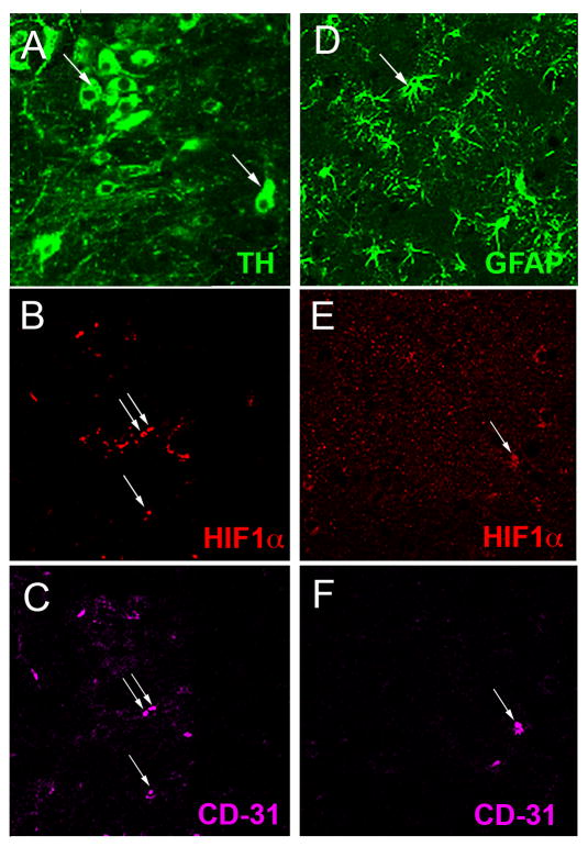
Fig. 2.
Cellular localization of HIF1α in the SNpc. (A–C): A section immunostained with TH (A, arrows), HIF1α (B, arrows) and the endothelial cell marker CD-31 (C, arrows) shows HIF1α present in endothelial cells but absent in TH+ DA neurons. GFAP+ astrocytes (D, arrow) are not labeled with HIF1α (E, arrow) that colocalizes with the endothelial marker CD31 (F, arrow).

