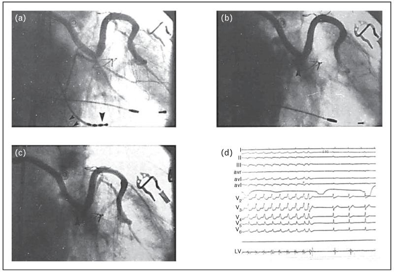
FIGURE 2.
Ethanol ablation of ventricular tachycardia. Right anterior oblique projection of the sequential vein graft. Panel a: Before the injection of ethanol, showing all three marginal branches of the circumflex artery (small arrowheads indicate the tachycardia-related artery, whereas large arrowhead is the mapping catheter localized on the site of origin of the tachycardia). Panel b shows immediately after the injection of ethanol, there is total occlusion of the circumflex (arrowhead). Panel c shows a 14-day follow-up angiogram showing persistent occlusion of the circumflex with minimal recanalization of the third marginal branch (arrowhead). Panel d shows termination of ventricular tachycardia immediately after the injection of ethanol. Left ventricular bipolar recordings obtained from the distal two poles of the quadripolar mapping electrode, with the catheter placed at the site of origin of the ventricular tachycardia. Note the marked splitting of the potentials (from Brugada et al. [24]).

