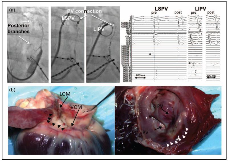
FIGURE 4.
Pulmonary vein isolation after ethanol infusion in the vein of Marshall (VOM). Panel a shows simultaneous LIPV and LSPV disconnection by VOM ethanol. Left image shows venograms with large VOM posterior branches, directed toward the LSPV and LIPV, and their reconnection sites (asterisks). Right image shows ethanol infusion led to disconnection of both veins. Panel b shows ethanol ablation lesion. Left heart shows epicedial aspect, showing the VOM and ligament of Marshall area, with pale discoloration of the ablated areas (arrowheads). Right heart shows endocardial aspect after incision in the left atrial appendage. A pale area of discoloration is shown anterior to the left pulmonary veins, surrounded by small area of tissue hemorrhage (arrowheads) (from Valderrábano et al. [44] and Dave et al. [45]).

