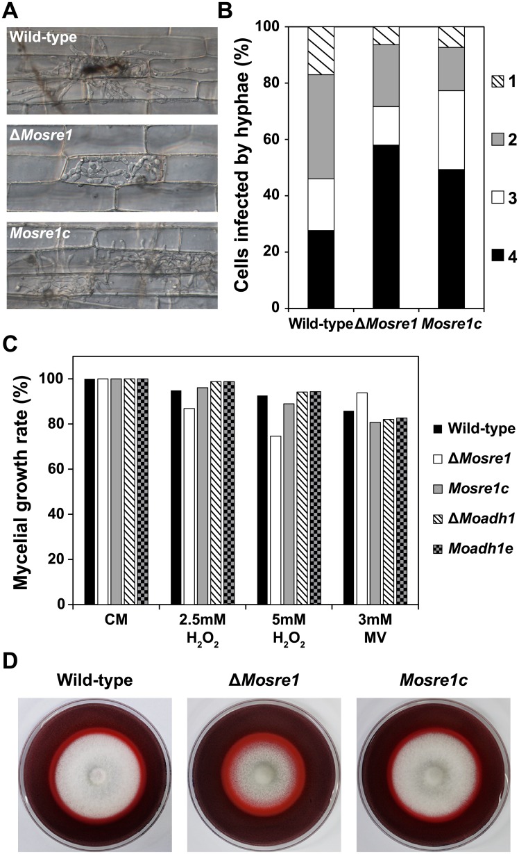
Fig 6. Invasive growth, oxidative stress sensitivity and enzyme activity of the wild-type, ΔMoadh1 and ΔMosre1.
(A) Infectious growth was observed in rice sheath cells. A conidial suspension (2 × 10⁴ conidia/ml) was inoculated into the excised rice sheath. Photographs were taken 48 hours after incubation. Scale bar indicates 20 μm. (B) Frequency of infected rice cells was determined by counting at least 100 appressorium-mediated penetration pegs with three replicates. Invasive growth was observed as described above. 1, Move to adjacent cell; 2, One cell filled; 3, Primary hyphae; 4, No penetration. (C) Extracellular oxidative stress sensitivity of the wild-type and two deletion mutants were examined. Wild-type and two deletion mutants were inoculated on CM and CM including 2.5 or 5 mM H2O2 and 3 mM methyl viologen (MV). (D) Wild-type, ΔMosre1 and Mosre1c were inoculated on CM containing 200 ppm Congo Red. Discoloration (halo) of Congo Red was observed at 9 days after incubation.

