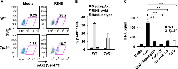
Fig 4. ERK and Akt are involved in Tpl2-dependent IFNλ production in pDCs.
pDCs from WT and Tpl2 -/- mice were stimulated with R848 for 18 hr, and analyzed by intracellular staining for pAktSer473. (A) Representative flow cytometry plots showing pAktSer473 staining within pDCs. (B) Proportion of pAkt positive pDCs from 2 independent experiments. (C) pDCs were pretreated with inhibitors for 30 min before stimulation with CpG, and IFNλ levels were measured by ELISA. Data are representative of 2 (A-B) or 3 (C) independent experiments. Graphs show means±SD. * indicates p<0.05, ** indicates p<0.01.

