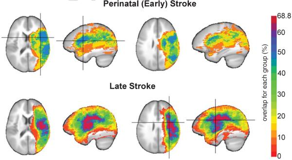
Fig. 1.
Lesion overlap maps in patients who suffered perinatal (early; n = 19; top) or late (n = 32; bottom) LMCA stroke. Stroke lesions are represented as the percentage of subjects who showed overlap in a region for each group and overlaid on to the ICB452 anatomical image. Crosshairs indicating the region of maximum overlap in perinatal stroke subjects (47.4%) was located in the left insula and in late stroke subjects (68.8%) was located in the left precentral gyrus.

