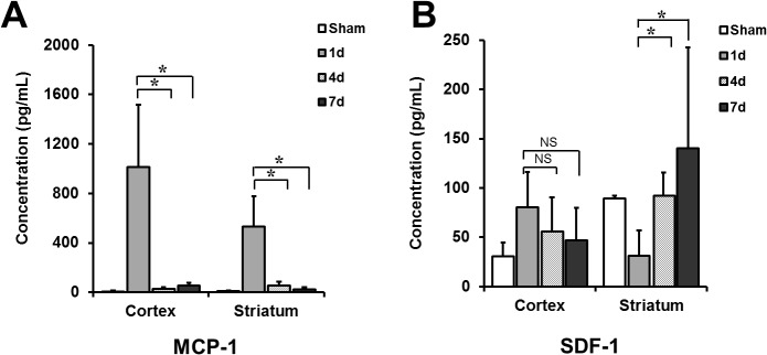
Fig 5. MCP-1 and SDF-1 expression levels in ischemic lesions according to the time after MCAo.

(A) Expression level of MCP-1 was higher in brain extracts obtained from both cortex and striatum at 1 day after MCAo than in those at 4 or 7 days after MCAo (*P < 0.01). (B) In comparison, the SDF-1 concentration was higher at 4 or 7 days after MCAo than the concentration at 1 day after MCAo in striatum (*P < 0.01). However, there was no difference in the SDF-1 concentration according to the time from MCAo in cortex (n = 4 for each group). MCP-1 indicates monocyte chemotactic protein-1; SDF-1, stromal cell-derived factor-1; MCAo, middle cerebral artery occlusion.
