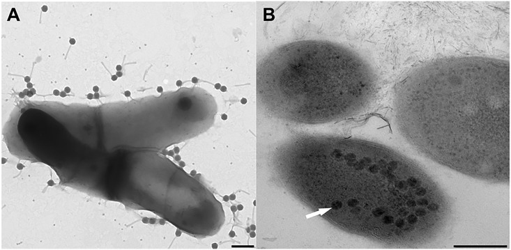
Fig 2. Stages in Gordonia phage infection cycles.
(A) Attachment stage of phage infection cycle between phage GMA6 and host Gordonia malaquae strain CON67. Scale = 200 nm. (B) Replication of phage GTE6 inside G. terrae strain CON34 cells prior to cell lysis. Arrows indicate phage replicated inside bacterial cells. Scale = 200 nm.

