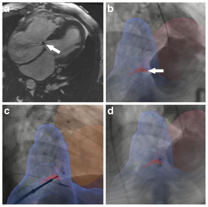
Fig. 1.
Clinical X-ray fused with MRI (XFM)-guided closure of ventricular-atrial (Gerbode) defect. a Four-chamber cine MRI showing defect between left ventricle and right atrium (arrow). Left-to-right flow is clearly seen. b XFM image in which the defect appears as a red overlay on the fluoroscopy (arrow). c Nitinol closure device on its delivery cable positioned across the defect. Both discs have been deployed. d Closure device after release. (Courtesy of Kanishka Ratnayaka (2014), Department of Cardiology, Children’s National Hospital Center, Washington DC, USA)

