Fig. 1.

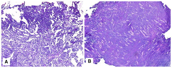
Histopathologic representation of BT specimens illustrating (a) TA-affecting tumor cells, causing moderate diagnostic difficulty, and (b) most of the sampled BT is unrecognizable due to TA (H&E staining, original low magnification)

Histopathologic representation of BT specimens illustrating (a) TA-affecting tumor cells, causing moderate diagnostic difficulty, and (b) most of the sampled BT is unrecognizable due to TA (H&E staining, original low magnification)