
Figure 3. Microengineered normal human mammary duct.
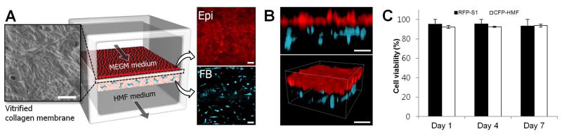
A. As visualized by scanning electron microscopy, the intervening vitrified membrane consists of a dense network of collagen fibers. This membrane supports adhesion and growth of mammary epithelial cells (shown in red; upper right inset) to a confluent monolayer, as well as 3D culture of fibroblasts (shown in cyan; lower right inset) within the collagen gel in the lower chamber. Epi and FB represent epithelial cells and fibroblasts, respectively. The micrographs were taken at day 7. Scale bars: 20 μm. B. A cross-sectional view (upper) and 3D rendered image (lower) show an intact normal mammary epithelium and fibroblasts embedded in the stromal layer of the lower chamber. Scale bars: 100 μm. C. In this microfluidic device, percent viability of the mammary epithelial cells (RFP-S1) and mammary fibroblasts (CFP-HMF) is maintained over 85% throughout the culture period (7 days). Data shown mean ± std.