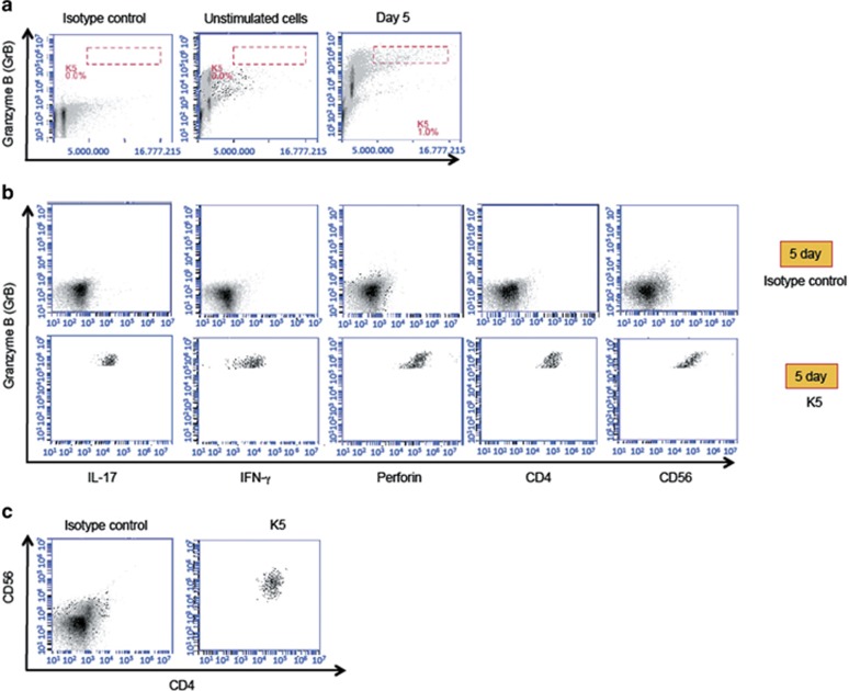
Figure 7.
Expression of surface markers and intracellular proteins in B19-specific PBMC. (a) PBMC from a subject ‘K' were incubated for 5 days with B19-VLPs (left and right panels) or with media alone (middle panel). Then B19-specific cells with highest intracellular GrB signal and forward scatter were gated (gate K5, right panel) for further analysis. Such cells were absent in unstimulated cells stained with GrB-antibody (middle panel) or B19 stimulated cells stained with GrB-isotype control antibody (left panel). (b) IL-17, IFN-γ, perforin, CD4 and CD56 expression signals in K5-gated cells are shown. (c) Left panel: total ungated PBMC stimulated with B19 VLPs and stained with isotype controls for CD4 and CD56 antigens. Right panel: co-expression of CD4 and CD56 antigens in B19-specific PBMC in gate K5.

