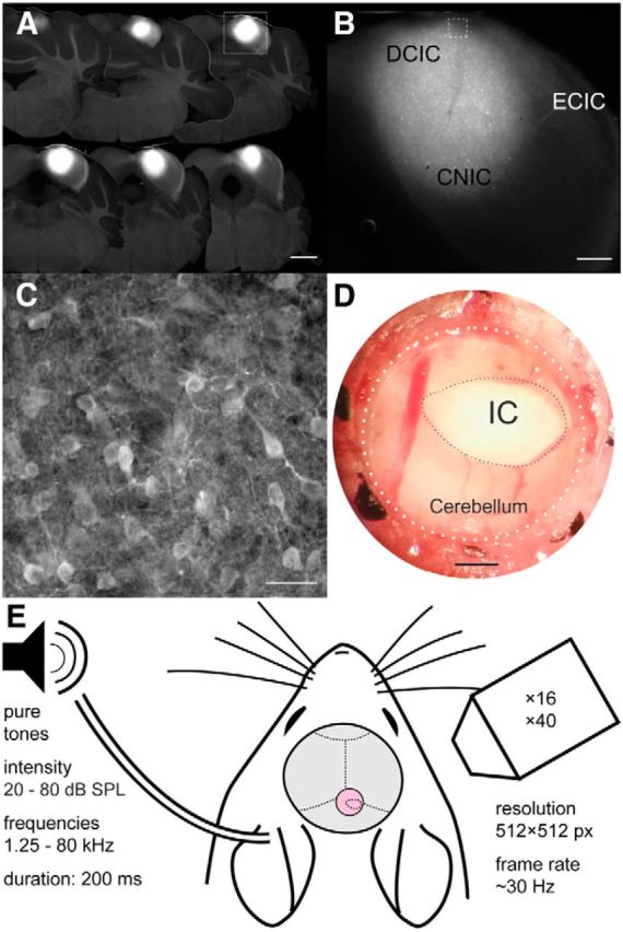
Figure 1.

GCaMP6m transduction of IC neurons and experimental setup. A, Coronal sections at 150 μm intervals showing widespread transduction of neurons throughout most of the dorsal parts of the IC. Scale bar, 1 mm. B, Higher-magnification image of one of the sections (A, white box) with the approximate locations of the DCIC, CNIC, and external cortex of the IC (ECIC) marked. Scale bar, 300 μm. C, Higher-magnification image (B, white box) showing individual IC cell bodies. Scale bar, 25 μm. D, Photograph of glass window (outlined by dotted lines) inserted into the skull over the IC. Scale bar, 500 μm. E, Experimental setup used for sound delivery and two-photon calcium imaging.
