
Figure 6.
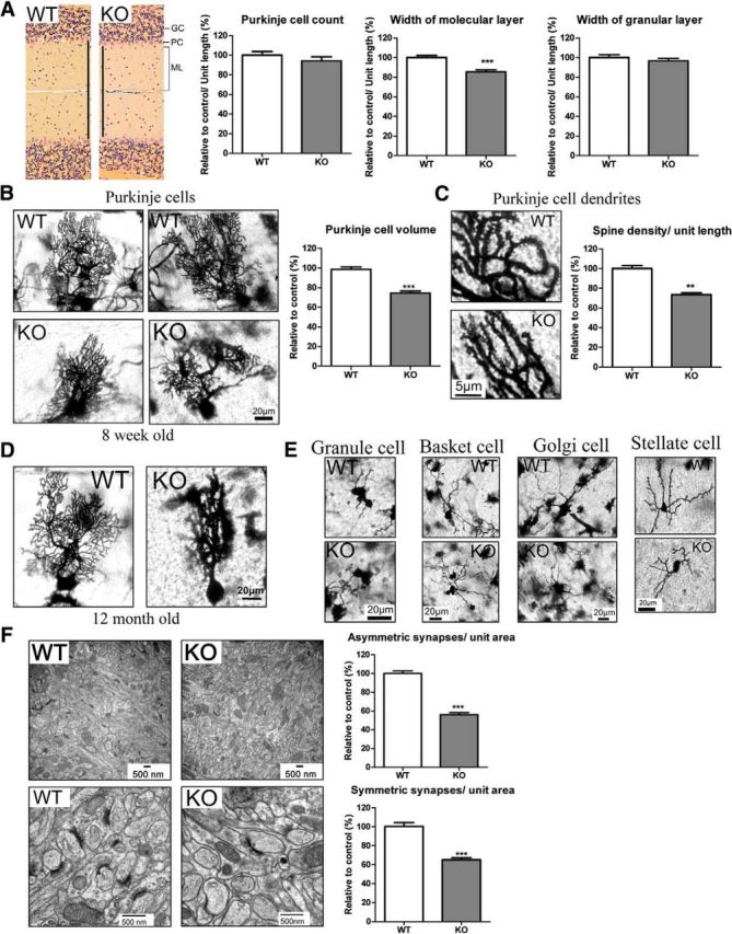
IP6K3 KO mutant mice manifest altered Purkinje cell structure and reduced synapses. A, H&E staining in cerebellum from wild-type and IP6K3 KO mice (8 weeks old). Width of the molecular layer is decreased in IP6K3 KO mice. The Purkinje cell number and the width of granule layer do not show any significant difference. B, Golgi staining reveals withered dendritic trees of Purkinje cells from 8-week-old IP6K3 KO mice cerebellum. Cell volume is significantly decreased in IP6K3 KO Purkinje cells. C, Golgi staining for the Purkinje cell dendrites from wild-type and IP6K3 KO mice cerebellum (8 weeks old). Spine number is significantly decreased in IP6K3 KO Purkinje cells. D, Golgi staining reveals withered dendritic trees of Purkinje cells from 12-month-old IP6K3 KO mice cerebellum. E, Golgi staining of granule cells, basket cells, Golgi cells, and stellate cells reveals shorter dendritic length (22 ± 5% less for basket cell, 15 ± 4% less for Golgi cell, and 25 ± 8% less for stellate cell) but no significant morphological change in the IP6K3 KOs (8 weeks old). We quantified the length of an average of five dendrites per cell, six cells per mouse. F, Electron microscopy of the cerebellar molecular layer from wild-type and IP6K3 KO mice (8 weeks old). Densities of both symmetric and asymmetric synapses are markedly decreased in IP6K3 KOs. The ultrastructure of synapses is relatively normal in the KOs. Data are represented as mean ± SEM, three mice per group, **p < 0.01, ***p < 0.001.