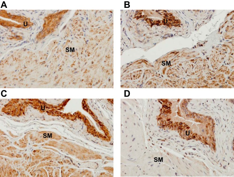
Fig. 5.
Representative photomicrographs showing immunohistochemical staining of MnSOD in transverse sections of bladder specimens from wild-type and Sod2lox/lox,SM-CreERT2(ki)Cre/+ mice, with or without OHT treatment, confirming that OHT treatment depleted MnSOD in detrusor SM of inducible SM-specific Sod2 deletion mice (modification: ×40). Bladder sections from a wild-type mouse without OHT injection (A), an OHT-treated wild-type mouse (B), and a Sod2lox/lox,SM-CreERT2(ki)Cre/+ mouse without OHT injection (C) show positive staining of MnSOD in detrusor SM and urothelium. Conversely, a bladder section from an OHT-treated Sod2lox/lox,SMCreERT2(ki)Cre/+ mouse (SM-specific Sod2−/−) (D) shows positive staining of MnSOD in urothelium, but not in detrusor SM. U, urothelium.

