
Fig. 8.
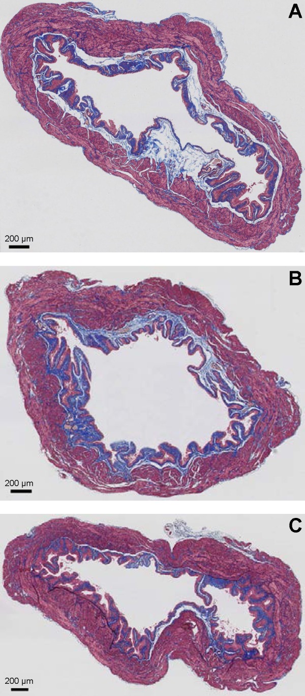
Representative images of Masson's trichrome staining of equatorial bladder sections of age-matched wild-type mice (A), SM-specific Cre mice (B), and SM-specific Sod2−/− mice (C) revealing SM (outer magenta), collagen (blue), and urothelium (inner light magenta). Scale bar indicates 200 μm.