Fig. 3.
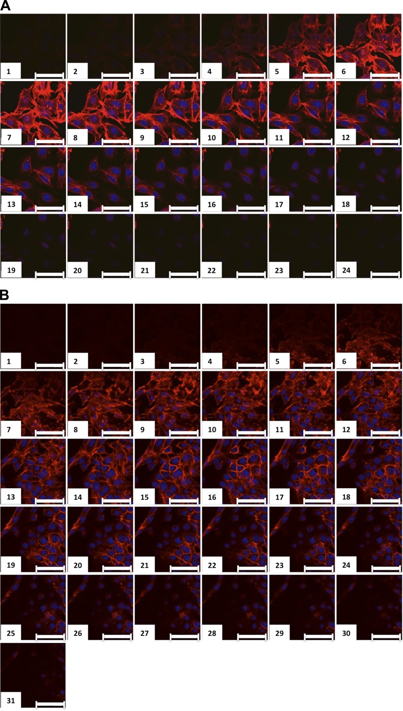
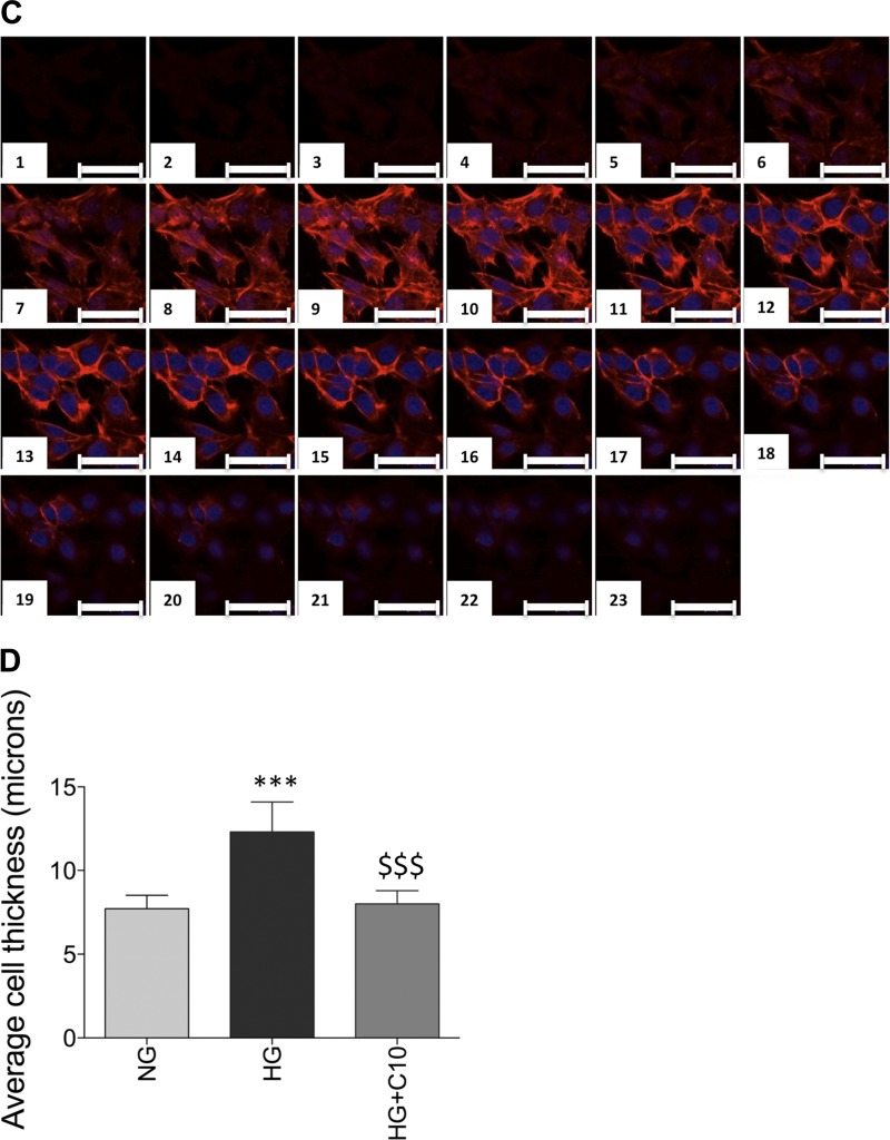
High glucose induces mesangial cell hypertrophy, which is reversed by glucosylceramide synthase inhibition. Mesangial cells were grown in normal glucose (NG; A), high glucose (HG; B), or HG supplemented with 0.15 μM C10 (C). At 48 h, cells were fixed, permeabilized, and stained with the F-actin marker phalloidin and the nuclear stain DRAQ. Serial 0.5-μm-thick z-sections were obtained using confocal microscopy. D: average thickness of mesangial cells calculated by the distance from the top to the bottom of the F-actin staining. Data are means ± SD; n ≥ 5. ***P ≤ 0.0001, HG compared with NG; $$$P ≤ 0.0001, HG + C10 compared with HG (as determined by one-way ANOVA). Scale bars = 50 μm.


