Fig. 1.
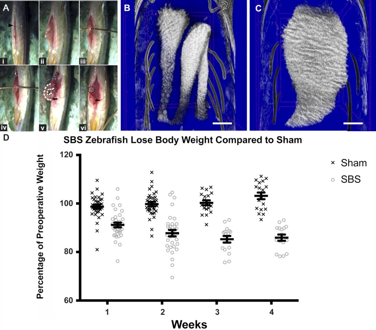
Zebrafish with short bowel syndrome (SBS) lose significantly more weight compared with sham-operated fish. A: SBS operation. i: Ventral laparotomy incision (solid black arrowhead). The head of the fish is located at the top of the image, while the tail is oriented at the bottom. ii: segments 2 and 3 (S2 and S3) are delivered through the incision. iii: A 22-gauge copper wire inserted beneath the bowel loop prevents the intestine from retracting back into the abdomen. iv: One wall of the proximal intestine is sutured to skin with a blue stitch. v: The proximal intestine is held in place along the abdominal wall by the suture (hollow arrowhead). The midportion of intestine (white dotted line) is resected and the distal intestine is suture ligated (black arrow). vi: The distal intestine (black arrow) is reduced into the abdomen leaving behind proximal S1 as an ostomy (dotted circle). B: micro-CT intestine following sham operation with proximal intestine at the top and distal intestine at the bottom of the image. Sham intestine is intact with normal caliber. C: micro-CT of intestine 2 wk after SBS surgery. The ostomy is present at the bottom of the image with the proximal intestine profoundly dilated. Scale bar = 500 μm. D: percentages of preoperative weight were plotted at each weekly time point with the surviving fish euthanized at 2 and 4 wk. Zebrafish with SBS lose a significant amount of weight compared with sham-operated fish. Error bars indicate SE.

