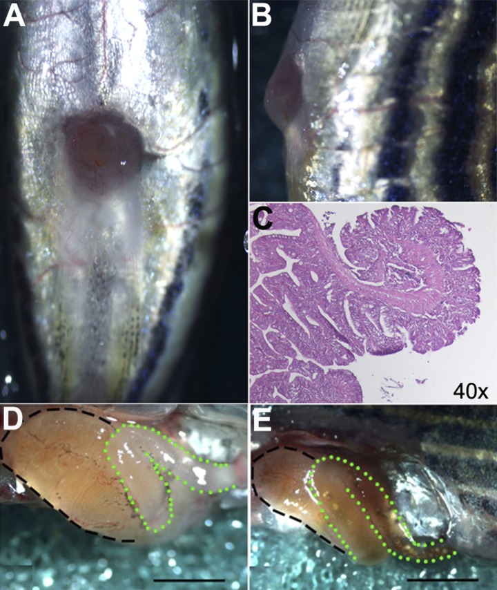
Fig. 2.
SBS fish demonstrate open and functional stomas and dilated proximal intestine at the time of euthanasia. A: ventral view demonstrates a patent ostomy and healed midline incision. B: stoma protrudes past the level of the skin on lateral view. C: hematoxylin and eosin stain on sagittal sectioning demonstrates the epithelial edge curling outward toward the skin. D: SBS fish demonstrate enlargement of the proximal intestinal segment S1 (black dashed line). The remaining distal intestine (green dotted line) is devoid of luminal content and feces, indicating that the distal intestine remains ligated and nonfunctional. E: sham-operation fish demonstrate a normal-appearing proximal segment (black dashed line) in continuity with distal intestine (green dotted line) evidenced by luminal content and feces from proximal segment to anus. Scale bar = 1 mm.

