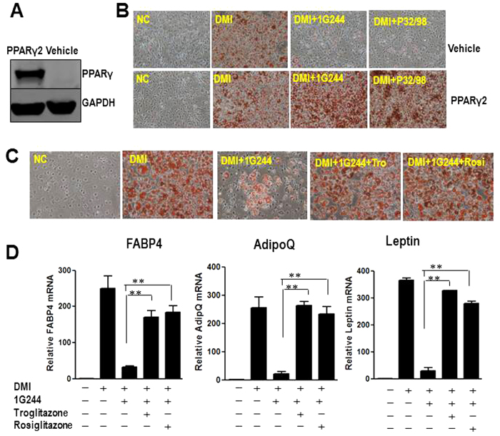
Figure 5. TZDs or ectopic PPARγ2 rescues inhibition of DPP8/9 induced adipogenic defects in 3T3-L1 cells.
(A) Representative western blot for the expression of PPARγ in stable 3T3-L1 cells transduced with control plasmid (vehicle) or PPARγ2 plasmid (PPARγ2). The blots were cropped, and the full-length blots are presented in the supplementary information. (B) Oil Red O staining of control cells or PPARγ2 overexpressed 3T3-L1 cells, treated vehicle (NC), DMI (DMI), 500 μM non-selective DPP4 family inhibitor P32/98 (DMI+P32/98) or 20 μM DPP8/9 inhibitor 1G244 (DMI+1G244) at day 8 of differentiation. (C) Oil Red O staining of 3T3-L1 cells treated with 20μM DPP8/9 inhibitor 1G244 (DMI+1G244) or 1G244 plus 1 μM rosiglitazone(DMI+1G244+Rosi) or 5 μM troglitazone (DMI+1G244+Tro). (D) Adipocyte markers, FABP4, adiponectin (AdipoQ) and leptin, were measured in these cells by real time PCR at day 8 of differentiation. β-actin expression was used as an internal control.

