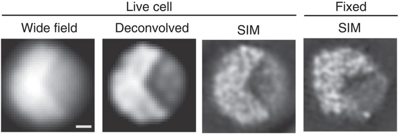
Figure 1. Appearance of chromatin in the interphase nucleus of S. pombe.
Chromatin structure in the interphase nucleus of S. pombe expressing H2B-GFP observed using 3DSIM. Single optical sections are shown. Raw (wide field), deconvolution and 3DSIM images of a single nucleus of a live cell are shown, together with a single 3DSIM image of another fixed cell. The contrast of the images is adjusted to show the background with a very low noise level. Fixed cells showed chromatin structures similar to the live cells. Scale bar, 500 nm.

