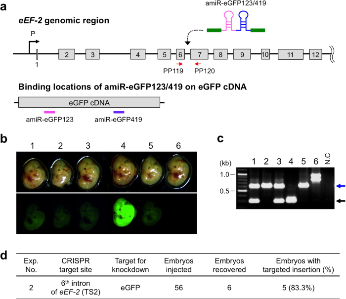
Figure 2. Targeted insertion of ssDNA encoding anti-eGFP amiRNA by CRISPR/Cas9 system (Exp. 2).
(a) Schematics of targeted integration of amiR-eGFP123/419 sequences into the intron 6 of eEF-2 gene (upper panel) and location of amiR-eGFP123 and -eGFP419 target sites on eGFP cDNA (lower panel). From the correctly targeted eEF-2 locus, amiRNAs get transcribed and subsequently bind to the target sites (amiR-eGFP123 = pink line and amiR-eGFP419 = purple line) on eGFP mRNA, leading to eGFP knockdown. Red arrows indicate the location of primer set (PP119/PP120) used for detection of fetuses with targeted insertion. (b) eGFP fluorescence among E13.5 day fetuses observed under a fluorescent stereomicroscope showing successful knockdown in some fetuses (see text for details). (c) Genotyping of fetuses by PCR using primer set shown in (a). The embryo numbers in (b) and (c) correspond with each other. Expected fragment sizes: wild-type = 301-bp (black arrow), targeted insertion = 625-bp (blue arrow). (d) Targeted insertion efficiency. Fetuses containing functional amiRNA sequence were considered as ‘embryos with targeted insertions’ even though insertions were not fully accurate.

