FIG 2.

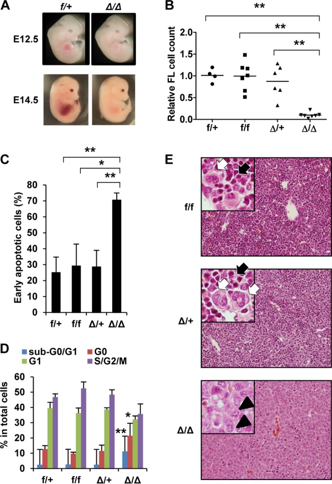
Fetal liver (FL) hematopoiesis is defective in Vav-iCre+ Srsf2flox/flox embryos. Vav-iCre+ Srsf2flox/+ mice were crossed with Srsf2flox/flox mice to generate Vav-iCre− Srsf2flox/+ (f/+), Vav-iCre+ Srsf2flox/+ (Δ/+), Vav-iCre− Srsf2flox/flox (f/f), and Vav-iCre+ Srsf2flox/flox (Δ/Δ) embryos. (A) Macroscopic appearance of E12.5 and E14.5 embryos. Δ/Δ embryos have smaller FLs than the controls. (B) Relative cell numbers of E14.5 fetal livers. Each cell number was normalized to the average of the numbers of f/+ FLs in the same litter. Horizontal bars show the average for each group. f/+, n = 4; f/f, n = 7; Δ/+, n = 6; Δ/Δ, n = 7. Δ/Δ FL cells were significantly fewer than those in other groups. (C) Apoptosis assay. Early apoptotic cells were defined as annexin V+ 7AAD− cells. f/+, n = 5; f/f, n = 2, Δ/+, n = 6; Δ/Δ, n = 6. Δ/Δ FLs had significantly more apoptotic cells than those in other groups. (D) Cell cycle analysis by pyronin Y and 7AAD staining. Δ/Δ FL cells show both a significant increase in the levels of apoptotic (sub-G0/G1) cells and a decrease in the levels of cycling (S/G2/M) cells compared to other groups. f/+, n = 5; f/f, n = 2; Δ/+, n = 6; Δ/Δ, n = 6. (E) Histology of E14.5 FL (hematoxylin-eosin stain). Δ/Δ FLs consist of hepatoblasts characterized by a large, pale-staining nucleus with distinct nucleoli (arrowheads). FLs of other genotypes are filled with hematopoietic cells, the majority of which are erythroblasts (black arrows), with scattered white blood cells (white arrows). Original magnification, ×200. Insets, ×400. Objective lenses, UPlanFL 20×, 0.50 numerical aperture (NA), and UPlanFL 40×, 0.75 NA (both from Olympus, Tokyo, Japan). Images were acquired at room temperature using an Olympus BX51 microscope equipped with a DP71 camera and DP controller/DP Manager software (Olympus, Tokyo, Japan). *, P < 0.05; **, P < 0.005.
