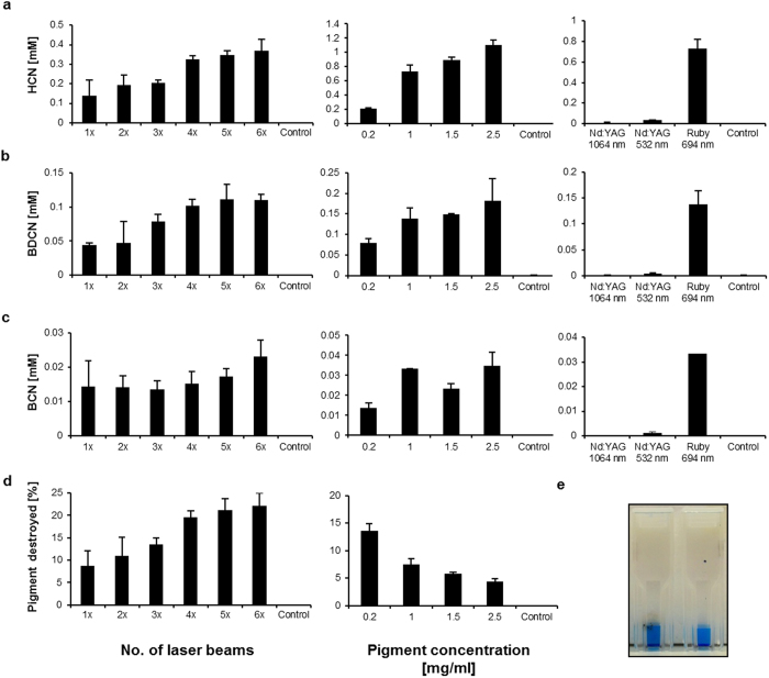
Figure 3. Phthalocyanine blue (pigment B15:3) is cleaved into BDCN, BCN and HCN upon laser irradiation.
(a)–(c) left: Levels of BDCN, BCN and HCN depending on the number of applied ruby laser pulses (1× to 6×; initial pigment concentration used: 0.2 mg/ml; control = no laser beam). (a)–(c) middle: BDCN, BCN and HCN levels as function of the initial pigment concentration (0.2 mg/ml to 2.5 mg/ml; at each concentration 3 laser pulses applied; control = no pigment). (a)–(c) right: Only slightly increased fragment concentrations were found upon Nd:YAG laser irradiation when compared to ruby laser irradiation (3 laser pulses at 1 mg/ml pigment; control = no laser beam). (d) left: Fraction (in %) of pigment destroyed depending on the number of ruby laser pulses applied (1× to 6×; initial pigment concentration used: 0.2 mg/ml; control = no laser beam). (d) right: Fraction (in %) of pigment destroyed depending on the initial pigment concentration (0.2 mg/ml to 2.5 mg/ml; at each concentration 3 laser pulses applied; control = no pigment). (e) UV/VIS cuvette after 3 laser pulses (left) compared to an untreated sample (right) containing 0.2 mg/ml of pigment B15:3 each. The laser-treated sample is carbonized at the outer cuvette surface but appears more intensely blue colored (see text for further details). All values are displayed as mean ± SD (n = 3).

