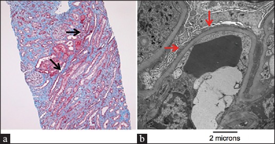
Figure 1.

Light and electron microscopic evidence of minimal change disease. (a) Light microscopy identifying focal interstitial fibrosis and cellular mesangial expansion (black arrows). No evidence of ischemic injury to tubular structures. Developed with periodic acid-Schiff stain. (b) Electron microscopic demonstration of widespread podocyte effacement (red arrows). Magnification, ×12,000
