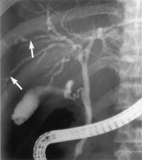
FIG. 2.
Posteroanterior endoscopic retrograde cholangiography in a 54-year-old man with clonorchiasis. Note the diffuse and uniform dilatation of the peripheral intrahepatic bile ducts with minimal dilatation of the extrahepatic bile duct and elliptical filling defects within the peripheral intrahepatic ducts, which correspond to the flukes (arrows).

