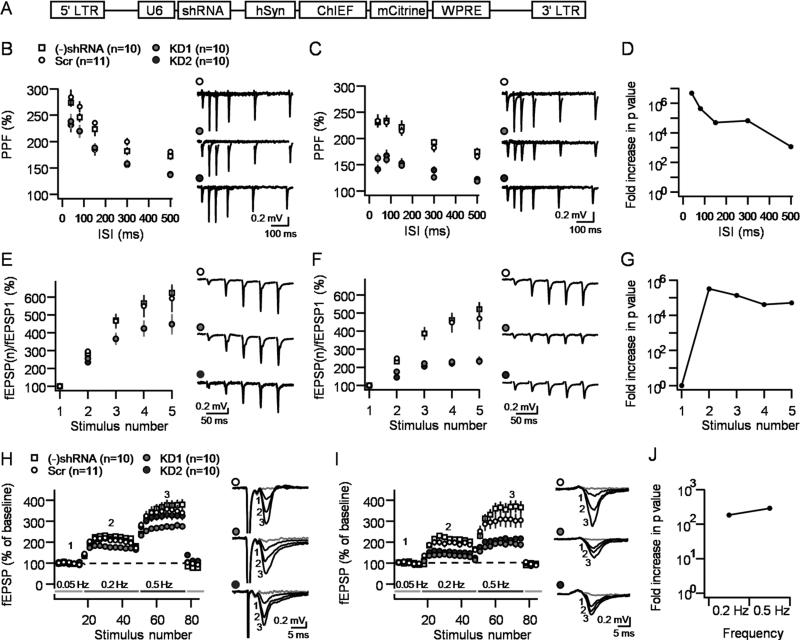
Figure 3. Optogenetic activation reveals major decrease in MF-CA3 short-term plasticity following tomosyn KD.
A. Diagram showing the genetic sequence packaged by the lentivirus used in this study. B,C. PPF of extracellular field potentials (fEPSPs) elicited by electrical (B) or optical (C) stimulation in the same slice under four conditions: naïve slices, non-targeting shRNA (Scr), KD1 and KD2. D. Dividing the p values of each ISI in the electrical stimulation by its optical counterpart reveals an increase of 3-7 orders of magnitude in significance levels. E,F. Reduced burst-induced facilitation in tomosyn KD slices under electrical (E) or optical (F). G. The statistical significance levels increased by more than five orders of magnitude for each stimulus in the burst. H,I. Reduced LFF in tomosyn KD slices observed under electrical (H) or optical (I) stimulation. Numbers in the representative traces correspond to numbers in the summary plots. J. The statistical significance levels increased by more than two orders of magnitude for each frequency step. Stimulus artifact was removed from traces in panels B and E for better visualization. fEPSP responses 10 minutes after DCG-IV application are denoted in gray. Number of slices for each condition is shown in brackets. Data are presented as mean ± S.E.M.

