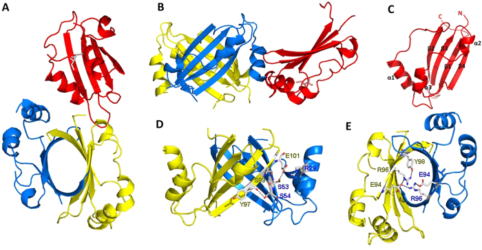
Figure 2. NosA1-111 overall fold.
(A): vertical view, (B): lateral view) Ribbon representations of a NosA trimer observed in an asymmetric unit, monomers were highlighted in red, blue and yellow, respectively. (C) monomer conformation, N-terminal and C-terminal and secondary structures were marked; (D) residues forming the hydrogen bonds in the β4 strand and the β2’ strand outside of β-barrel; (E) the salt-bridge and hydrogen-bonds formation between R96 and E94’ within the β-barrel.

