Figure 2.
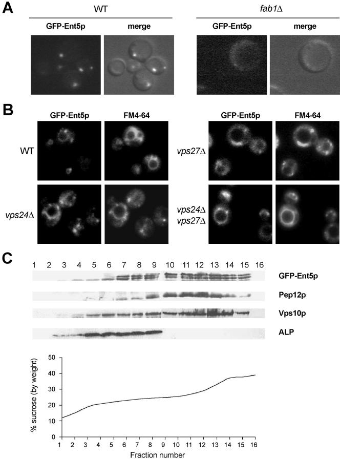
GFP-Ent5 localizes to endosomal structures. (A) Wild-type cells (FY833) and fab1Δ1 expressing GFP-Ent5 (pFL644) were observed by fluorescent and Nomarski microscopy. (B) Wild-type cells (BY4741), vps24Δ, vps27Δ, and vps24Δ vps27Δ cells expressing GFP-Ent5 (pFL644) cells were labeled with FM4-64 for 20 min to reveal endosomes and vacuolar membranes and observed by fluorescent microscopy. (C) ent5Δ cells (FLY675) expressing the GFP-Ent5 fusion construct were subjected to subcellular fractionation on a sucrose density gradient. High-speed membrane pellet (100,000 × g) from ent5Δ cells (FLY675) expressing the GFP-Ent5 construct was fractionated upon an equilibrium sucrose density gradient. Fractions were assayed by immunoblotting for GFP-Ent5, Pep12p, Vps10p, and ALP. Fractions 1-16 are shown. The immunoblots presented are representative of four separate experiments.

