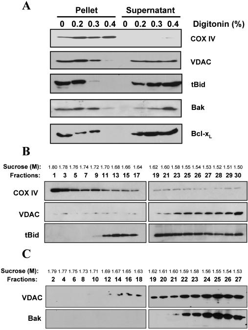
Figure 2.
Localization of tBid in the mitochondrial outer membranes. (A) Localization of tBid, Bak, and Bcl-xL in the mitochondrial outer membranes. Mitochondria were incubated with tBid (0.1 μg/ml) or Bcl-xL (13 μg/ml) for 1 h at 30°C before being treated with different amount of digitonin (wt/wt, 0-0.4%). Mitochondria were then separated by centrifugation. The supernatant, containing the outer membranes, and the pellet, containing the inner membranes, were analyzed by SDS-PAGE followed by immunoblot for the proteins indicated. (B) Distribution of tBid on different mitochondrial fractions. Mitochondria were incubated with tBid as in A, and then disrupted and fractionated on a sucrose linear gradient. Eluted fractions were analyzed by SDS-PAGE and immunoblot. (C) Mitochondria without tBid treatment were fractionated as in B and analyzed by immunoblot with antibodies against VDAC and Bak. For B and C, the sucrose concentration of each fraction is indicated.

