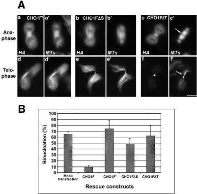
Figure 5.
ATP-binding mutant of CHO1 (CHO1F′) and CHO1 constructs lacking stalk (CHO1FΔS) or tail (CHO1FΔT) domains failed to rescue cytokinesis in endogenous CHO1-depleted cells. (A) CHO1F′ (a and d) and CHO1FΔS (b and e) constructs decorate the spindle microtubules throughout mitosis, but lack the ability to concentrate at the midzone/midbody region. In contrast to CHO1FΔT (c and f), CHO1F′ and CHO1FΔS constructs do not facilitate formation of the midbody matrix, based on continuous α-tubulin staining in the middle of the intercellular bridge (d′ and e′). CHO1FΔT localizes at the midzone (c) and midbody (f) and facilitates formation of the dense matrix (arrows in c′ and f′). Cells were stained with polyclonal anti-HA (a-f) and anti-α-tubulin (a′-f′) antibodies. Bar, 10 μm. (B) Histograms show no substantial decrease in the level of binucleation after rescue with CHO1F′, CHO1FΔS, and CHO1FΔT constructs.

