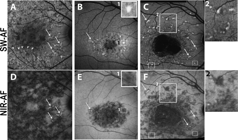
Figure 1.
Autofluorescent flecks viewed in short-wavelength autofluorescence (SW-AF) (A–C) and near-infrared autofluorescence (NIR-AF) (D–F) images of patient 16 (A, D), patient 4 (B, E), and patient 17 (C, F). Flecks can be widespread (A, D) or limited to perifoveal areas (B, E) and can be isolated (C, F) or in contact with one another (area in [A] indicated by arrowheads). Flecks identified as bright profiles in SW-AF images are most often dark in NIR-AF images (B, E; C, F). Flecks that exhibit SW hyperautofluorescence (arrows in [A–C]) can sometimes have central NIR-AF intensity (arrows in [D–F]). In SW-AF images, flecks can exhibit a dark halo ([C] inset 2). The area of bright SW-AF signal in flecks ([B] inset 1; [C] small rectangles) is typically smaller than the area of low NIR-AF of flecks ([E] inset 1; [F] small rectangles).

