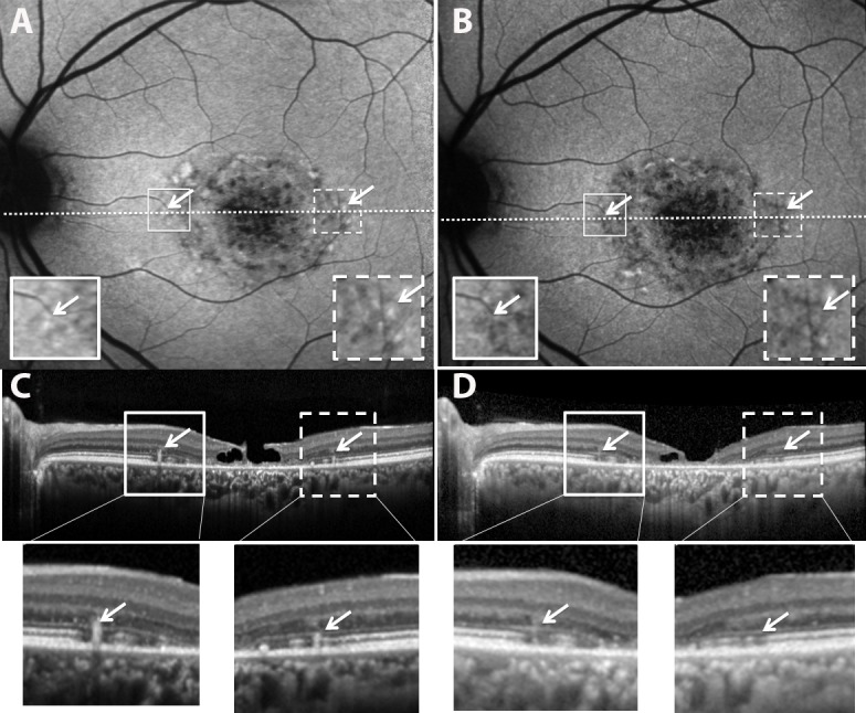
Figure 8.

Resorbing flecks in SW-AF and SD-OCT images (patient 1). Images in (B) and (D) were acquired 2.3 years after images in (A) and (C). Flecks having reduced brightness in the SW-AF image in (B) ([B] versus [A], arrows) also exhibit hyporeflectivity in the SD-OCT scan ([D] versus [C]). Expanded views of the outlined areas in (A) and (B) and shown as insets; expanded views of outlined areas in (C) and (D) are shown below.
