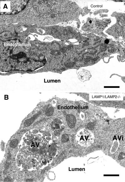
Figure 2.
Autophagic vacuoles accumulated in cells of LAMP-1/LAMP-2 double-deficient embryos. (A) Endothelium of a wild-type embryo. (B) Endothelium of a blood vessel in the neural tube in a LAMP-1/LAMP-2 double-deficient embryo. The endothelial cells displayed several cytoplasmic vacuoles with polymorphous contents. Some of the vacuoles could be identified as autophagic vacuoles (AV) or autophagosomes (AVi). Bars, 0.5 μm.

