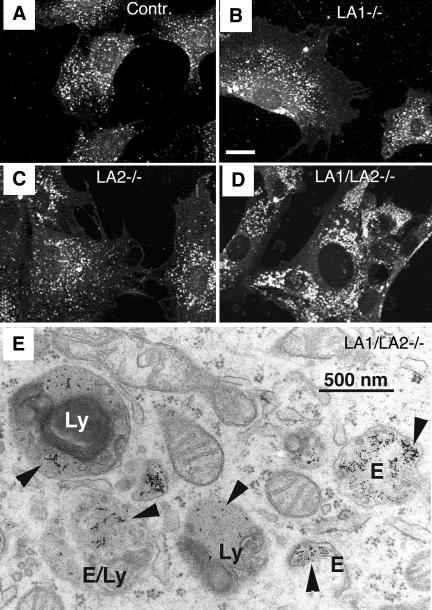
Figure 4.
The lysosomal compartment in LAMP double-deficient MEFs. Immunofluorescence staining of LIMP-2/LGP-85 in (A) control, (B) LAMP-1-deficient, (C) LAMP-2-deficient, and (D) LAMP-1/LAMP-2 double-deficient MEFs. Note the larger size and the more peripheral location of lysosomes in LAMP double knockout cells. Bar, 20 μm. The images show representative cells, based on experiments with at least two independent cell lines for each genotype. (E) Electron micrograph of LAMP-1/LAMP-2 double-deficient cells fed with the fluid-phase endocytic marker BSA-gold for 2 h. Structures with a typical morphology of dense lysosomes (Ly) or endosomes (E) contain the endocytic marker (arrowheads). Note also the multilamellar membranes inside the lysosome on the left.

