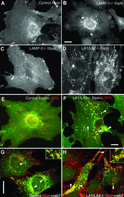
Figure 7.
Cholesterol accumulation in LAMP-1/LAMP-2 double-deficient MEFs. Filipin staining revealed distribution of unesterified cholesterol in (A) control, (B) LAMP-1-/-, (C) LAMP-2-/-, and (D) LAMP-1/LAMP-2-/- cells. Note increased vesicular staining in LAMP-2 single-deficient cells and prominent vesicular accumulation in LAMP double-deficient cells. (E and F) Double labeling with filipin (green) and the late endosomal marker LBPA (red) in control (E) and LAMP double-deficient cells (F). Yellow color (arrowheads) indicates colocalization in a subset of the endosomes of LAMP double knockout cells (F). (G and H) Double labeling of filipin (red) and GFP-rab7 (green) in control (G) and LAMP-1/LAMP-2 double-deficient cells (H). Rab7 colocalization in filipin positive vesicles is indicated by arrowheads and shown at higher magnification in the inserts. Bars, 20 μm.

