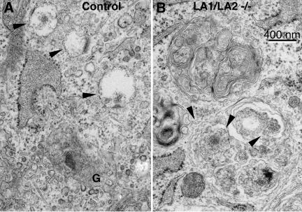
Figure 8.
Electron microscopical visualization of filipin labeled membrane cholesterol. Arrowheads in control (A) and double-deficient fibroblasts (B) indicate the filipin labeling. In control cells labeling was detected in the limiting membranes of small vesicles close to the Golgi apparatus (G). In LAMP-1/LAMP-2 double-deficient fibroblasts (B) filipin-induced membrane alterations were seen in both the limiting and internal membranes of endo/lysosomal vesicles. Note that lamellar internal membranes of the upper endo/lysosome are not labeled by filipin.

