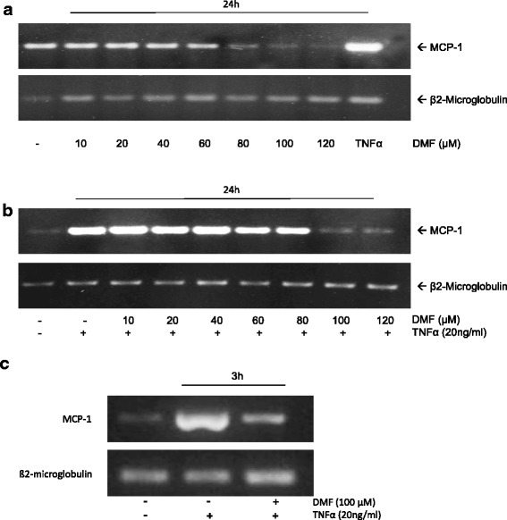
Fig. 2.

Analysis of constitutive and TNF-α-induced MCP-1 mRNA expression during the treatment with DMF in HUVECs. We isoalted total cellular mRNA and performed RT-PCR analyses for MCP-1 and β2-microglobulin. a HUVECs were mock-treated (solvent only) or treated with DMF at the indicated concentrations for 24 h. b HUVECs were mock-treated (solvent only) or treated with TNF-α (20 ng/ml) or DMF at the indicated concentrations + TNF-α for 24 h. c HUVECs were mock-treated (solvent only) or treated with TNF-α (20 ng/ml) or DMF (100 μM) + TNF-α for 3 h. The experiments were performed with comparable results at least 5 times
