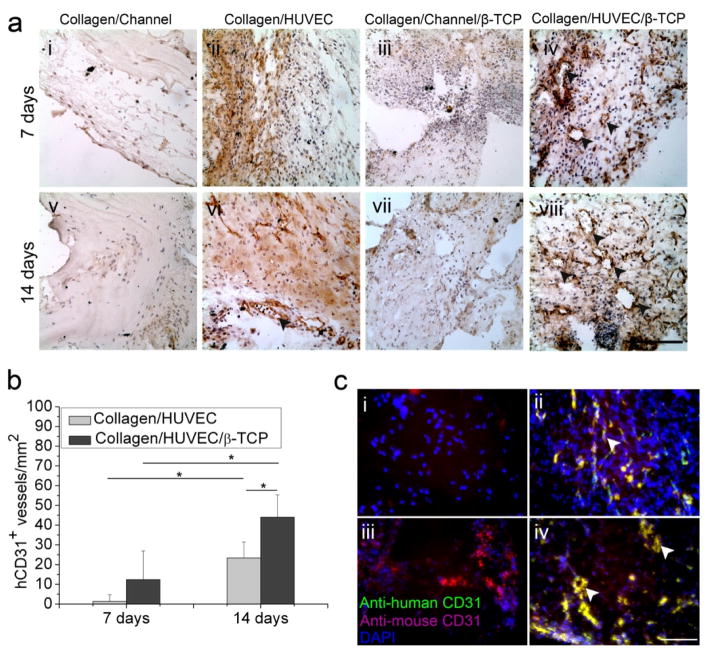
Fig. 5.
Immunohistochemistry evaluation of angiogenesis and anastomosis in vivo. (a) Representative immunohistochemistry images of human-CD31 (hCD31) from Collagen/HUVEC and Collagen/HUVEC/β-TCP grafts at day 7 and 14 (i-viii) showed that the hCD31 positive microvessels contain murine erythrocytes (iv,viii) (black arrowheads) (Scale bar=100 μm). (b) The hCD31-positive expressing lumens containing murine erythrocytes were quantified by measuring their density (*p<0.05, n=8). (c) Immunofluorescent staining of human CD31 (green) and mouse CD31 (magenta) shows the anastomosed sites of preformed human capillaries with host vasculature (white arrows, yellow color). Four groups at day 14 are shown: (i) Collagen/Channel, (ii) Collagen/HUVEC, (iii) Collagen/Channel/β-TCP, and (iv) Collagen/HUVEC/β-TCP.

