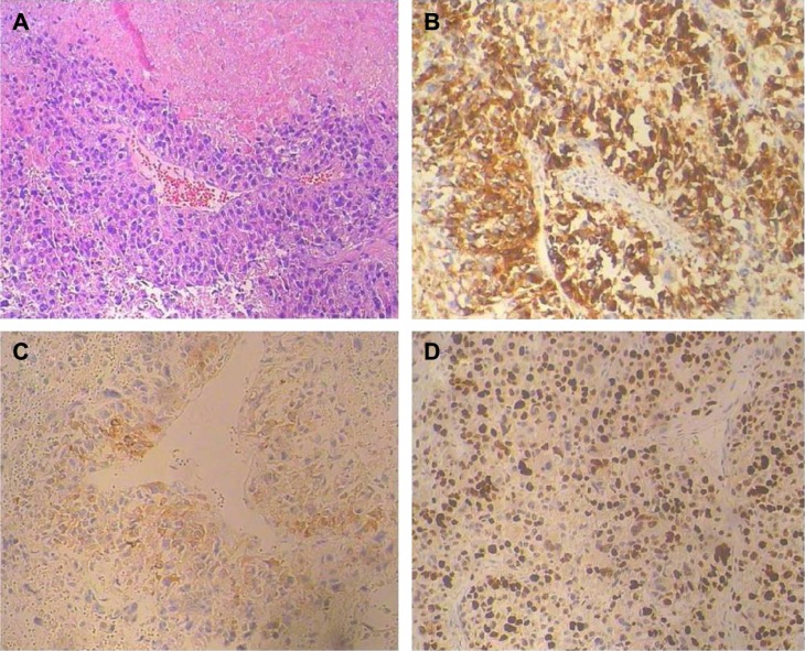
Figure 1.
Pathological findings of the pulmonary metastases from malignant uterine PEComa.
Notes: (A) Hematoxylin and eosin stain, magnification ×100. (B and C) HMB-45 and SMA immunohistochemical stain, magnification ×200. (D) Ki-67 immunohistochemical stain, magnification ×200. The average Ki-67 labeling index is 40% in this tumor. Background staining was identified by negative controls in which the sections were performed by substitution of primary antibodies with phosphate buffer solution.
Abbreviations: HMB-45, human melanoma black 45; PEComa, perivascular epithelioid cell tumor; SMA, smooth muscle actin.

