Figure 1.
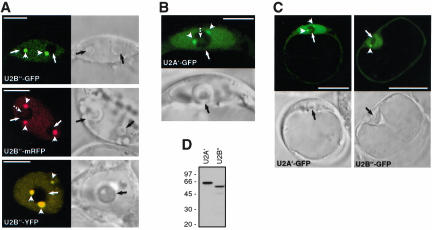
Analysis of FP-tagged U2 snRNP-specific proteins U2B″ and U2A′ in transiently transformed Arabidopsis protoplasts. (A) Localization of U2B″-GFP, U2B″-mRFP, and U2B″-YFP fusion proteins. Arrows and arrowheads point to nucleoli and Cajal bodies, respectively. Broken arrow points to the central nucleolar vacuole. (B) Localization of U2A′-GFP fusion protein. Arrows and arrowheads point to nucleoli and Cajal bodies, respectively. (C) U2A′-GFP and U2B″-GFP fusion proteins localize to nucleus and cytoplasm. Arrowheads point to Cajal bodies and arrows to nucleoli. Bars, 7 μm (A and B) and 25 μm (C). In A-C, single confocal sections with corresponding DIC images are shown. (D) Immunodetection of U2A′ and U2B″ GFP fusion proteins in protein extract from transformed protoplasts. Total protein extracts were analyzed by SDS-PAGE and Western blotting with anti-GFP antibody. Molecular mass standards in kilodaltons are indicated on the left.

