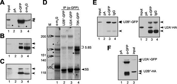
Figure 3.
Transiently expressed U1- and U2 snRNP-specific proteins assemble into mature snRNP. Immunoprecipitation of U2B″-GFP (A), U2A′-GFP (B), and U1-70K-GFP (C) fusion proteins. Lane 1, input protein extract. Lane 2, protein extracts incubated with protein A-Sepharose (pA). Lane 3, immunoprecipitations with anti-GFP antibody (α-GFP). Lane 4, immunoprecipitations with anti-m3G antibody (α-m3G). The blots are probed with anti-GFP antibody. Arrows in A-C point to U2B″, U2A′, and U1-70K-specific bands, and arrowheads point to immunoglobulin heavy chains. Bands marked with an asterisk in A are degradation products of U2B″-GFP. Only relevant portions of the blots are shown. (D) Analysis of anti-GFP immunoprecipitates for the presence of snRNAs. Lane 1, RNA immunoprecipitated with anti-m3G antibody. Lanes 2 and 3, RNA coprecipitated with U2B″-GFP and U2A′-GFP, respectively. Lane 4, RNA coprecipitated with U1-70K-GFP. Four spliceosomal snRNAs are indicated. Bands marked with asterisks are of unknown identity. Size markers in nucleotides are indicated on the left. 5S and 5.8S RNAs, which occur as background, are indicated on the right. (E) Coprecipitation of U2B″-GFP and U2A′-HA proteins. Protein extracts from protoplasts cotransformed with plasmids expressing U2B″-GFP and U2A′-HA were immunoprecipitated with anti-GFP antibody. Lane 1, input protein extract. Lane 2, protein extract incubated with protein A-Sepharose beads alone. Lane 3, immunoprecipitation with anti-GFP antibody. Lane 4, immunoprecipitation with unspecific antibody. The same blot was probed with anti-GFP (left) and after stripping anti-HA antibodies (right). (F) Coprecipitation of U2A′-GFP and U2B″-HA. Protein extracts from protoplasts cotransformed with plasmids expressing U2A′-GFP and U2B″-HA were immunoprecipitated with anti-GFP antibody. Lane 1, protein extract incubated with protein A-Sepharose beads alone. Lane 2, immunoprecipitation with anti-GFP antibody. Lane 3, input protein extract. Arrowheads in E and F point to immunoglobulin heavy and light chains.

