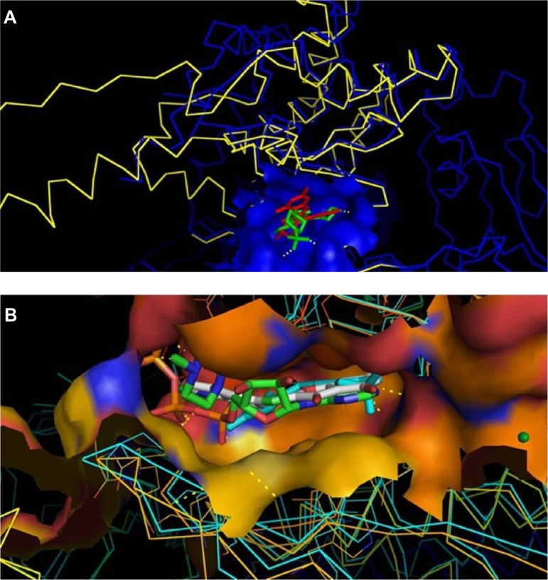
Figure 7.
Docking of known inhibitors into 3D model construction.
Notes: Panel (A) shows the model built with a docked ligand superposed at the template 3L13 and its ligand. The model is in yellow, the PI3K (3L13) in blue, docked ligand (WYE) in red and the PI3K crystallized ligand (JZW) was in green. Panel (B) shows the superposition of the model of the ligand docked into catalytic site to ATP crystallized PI3K (PDB ID: 1E8X). The docked ligand (WYE) is in white, the ATP complexed into 1E8X in green, the BYM complexed into 2A4Z in Cyan.
Abbreviations: PDB, Protein Data Bank; ATP, adenosine triphosphate.

